Paket 'Angiology anatomy' sadrži 30 naljepnica za dnevne razgovore. Besplatno preuzimanje za instalaciju na WhatsApp.
Fraze na naljepnicama:
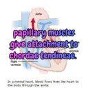
Aorta papillary muscles give attachment to ventricle chordae tendineae. Right ventricle In a normal heart, blood flows from the heart to the body through the aorta.
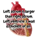
Left atrium is larger than right atrium. Left ventricle's wall is thickest of all.
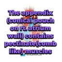
The appendix (conical pouch on rt. atrium wall) contains pectinate(comb ount Sinai Health System like) muscles De-oxygenated blood
Wt. of adult heart is 0.5% of body wt. Os cordis present in ruminanats.
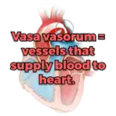
Pericardium is Aubunal aorta attached to phragm Vasa vasorume vessels that supply blood to heart.
moderator band/ septomarginal trebecula- prevent excess dilation of heart.
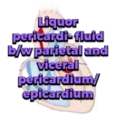
Liquor pericardi-fluid b/w parietal and viceral pericardium/ epicardium
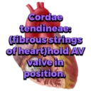
Cordae tendineae: (fibrous strings of heart) hold AV valve in position.
5/7 th portion of heart lies on left side because rt. lung is larger in size. Sternum aorta
Descending aorta- Pulmonary Circumfle left heart of fowl is Cranial venacava placed at cranial Right atrium part of thoraco- abdominal Right ventricle Left ventricle- cavity
Descending aorta- Pulmonary arteries- Pulmonary vein Caudal venacav Circumflex branch of left coronary artery Brachiocephalic trunk Pericardico- phrenic acava Right atrium -Right coronary artery ligament present in dog.
The common pulmonary trunk is connected to the descending aorta by fibrous cord called LIGAMENTUM ARTERIOSUM
Parts of vessels: tunica intima tunica media tunica adventitia
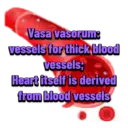
Vasa vasorum: vessels for thick blood vessels; Heart itself is derived from blood vessels
Anastomosis- communication b/w blood vessels, 2 types: inter-atrial and arterio-venous
End arteries-which do not form any pre-capillary anastomosis.
Descending aorta- Pulmonary arteries- Pulmonary veins. Caudal venacava Circumflex left corona Brachiocephalic trunk Cranial venacava In fowl, rt. AV valve is -Right huidm guarded by only one strong muscular leaf. Left ventricle-
Red Bond Cott aorta:2 parts ascending-beginning portion; decending caudal aortă
aortic arch-sharp curve of ascending aorta, whichpasses caudally and lead to descending aorta
aortic sinus/bulbus aorticus:it is the bulbus swelling on the wall of aorta just at the beginning
Red Blood Colly brachiocephalic. trunk:large vessel originate from aortic *arch at level of 4th rib and proceed cranially
brachiocephalic trunk detaches left and right axillary/subclavian artery
In dogs and pigs, left axillary/subclavian... artery arises directly from aortic arch and White Wood Cell not detached from brachiocephalic trunk
Close to thoracic inlet and ventral to trachea, the bicarotid trunk bifurcate intort. & left common carotid artery.
UMBILICAL ARTERY UMBILICAL VEIN UMBILICAL CORD BLOOD in foetal stage, 2 umbilical arteries and 1 umbilical vein (left BLICAL vein)are present AMNION WHARTON'S JELLY TISSUE
UMBILICAL VEIN UMBILICAL CORD BLOOD umbilical vein carry oxygenated blood towards liver. umbilical artery carry UMBILICAL ARTERY deoxygenated blood. WHARTON'S JELLY TISSUE
Ductus arteriosus connect pulmonary artery with desc. aorta. After birth, it occluded to form ligamentum arteriosus. e arteries
Foramen ovale-connects rt. atrium with left atrium to pass max. blood. Hypogastre arteries
umbilical vein join with: 1.portal vein 2 hepatic vein 3.ductus venosus 4.abdominal vena cava 5.rt.atrium
kajal(vet bhai ka gyan)
28-12-2023

